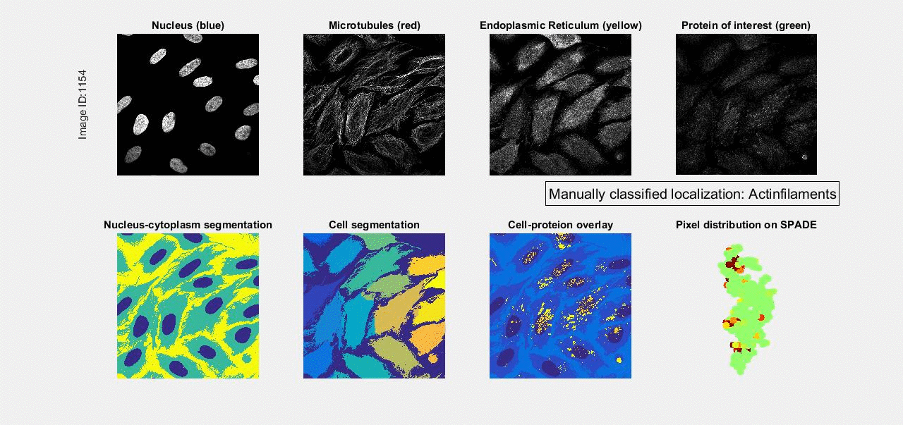
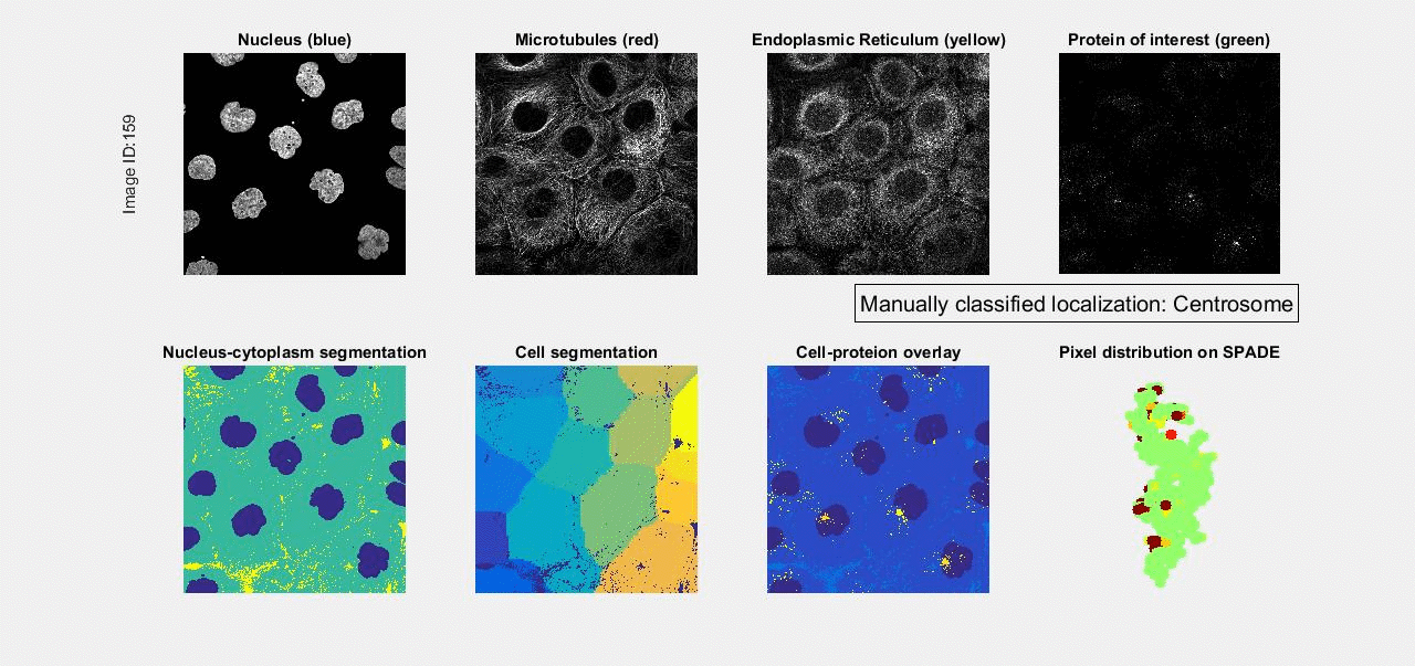
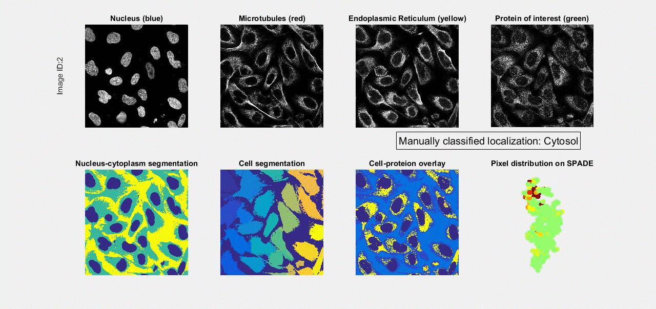
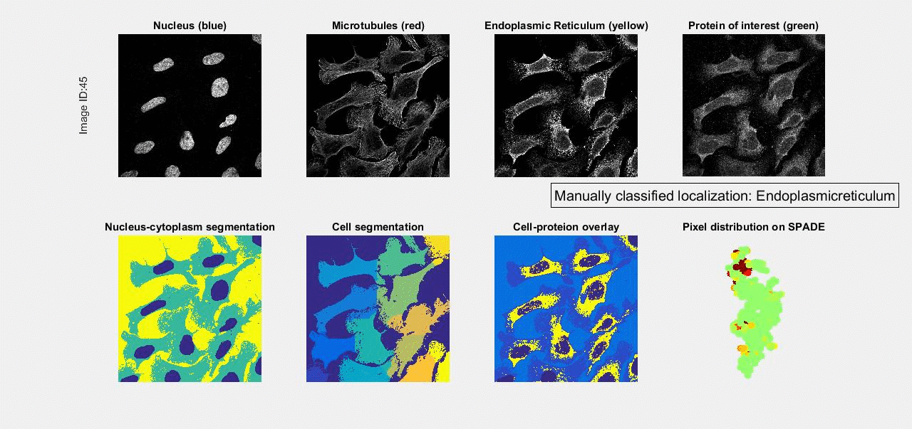
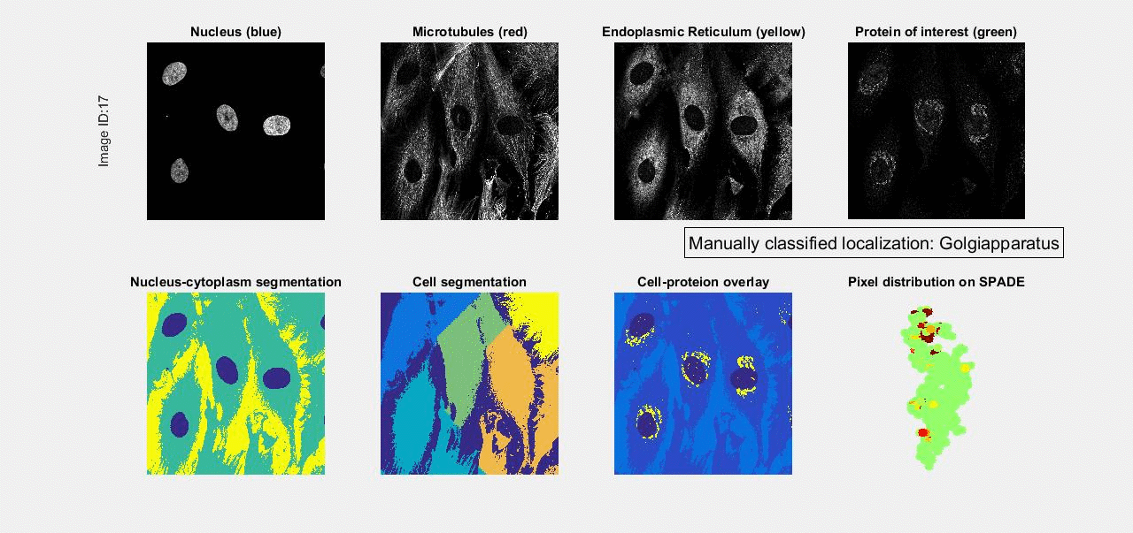
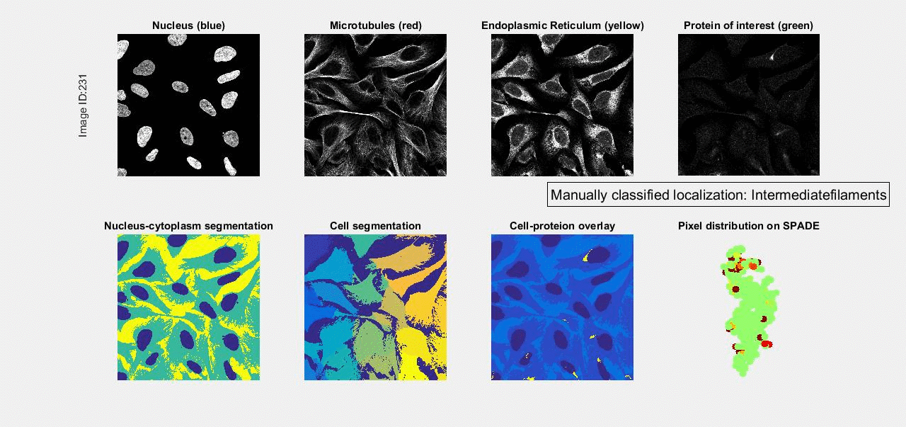
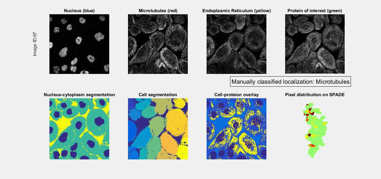
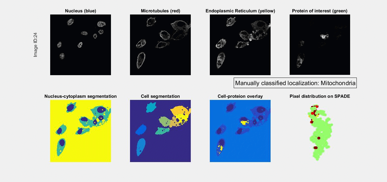
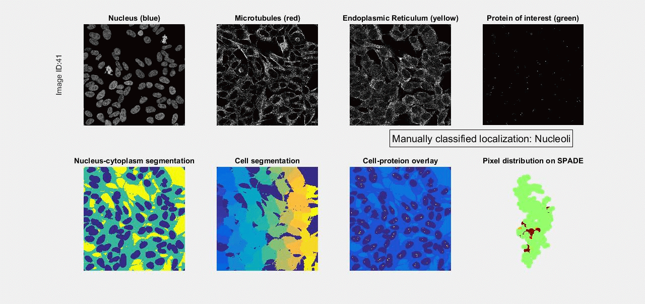
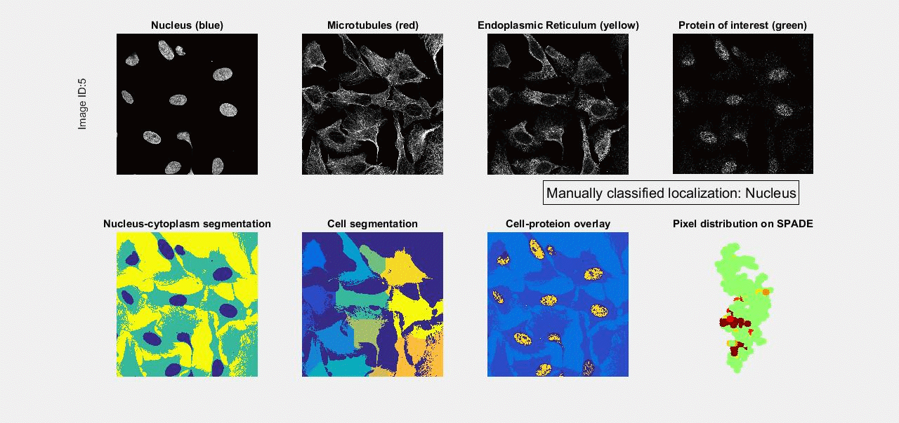
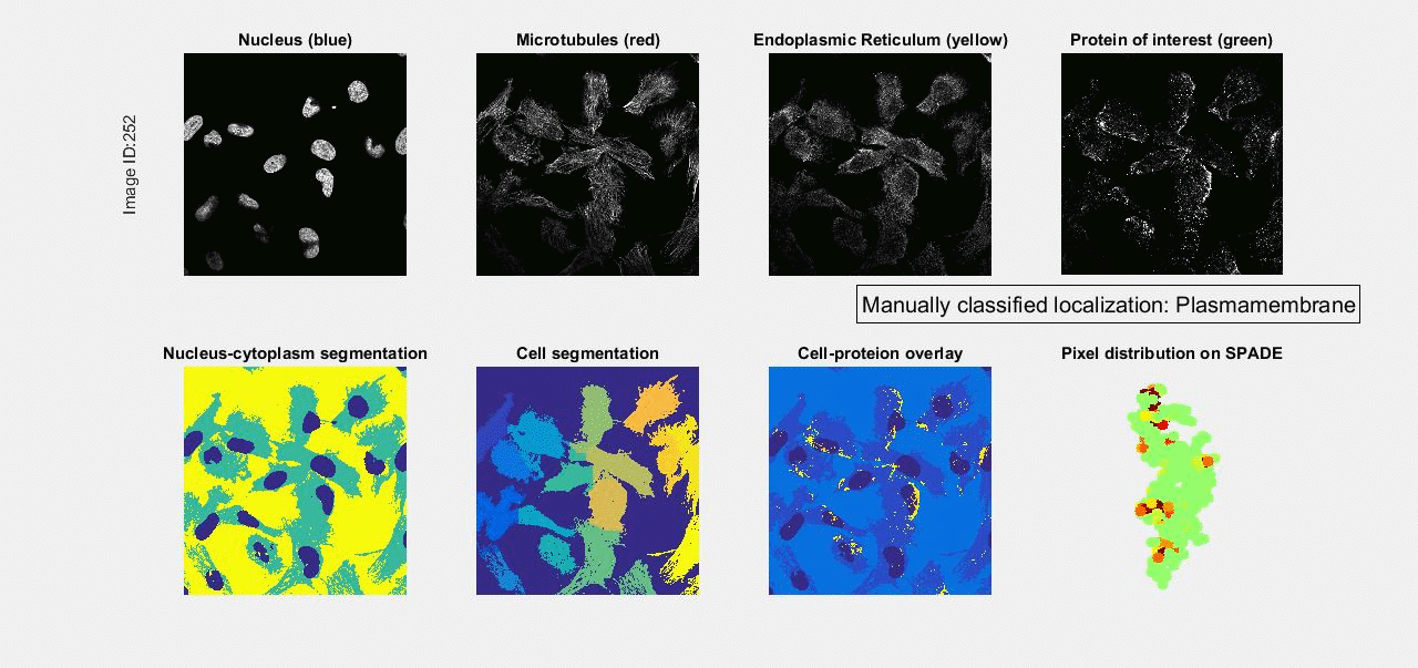
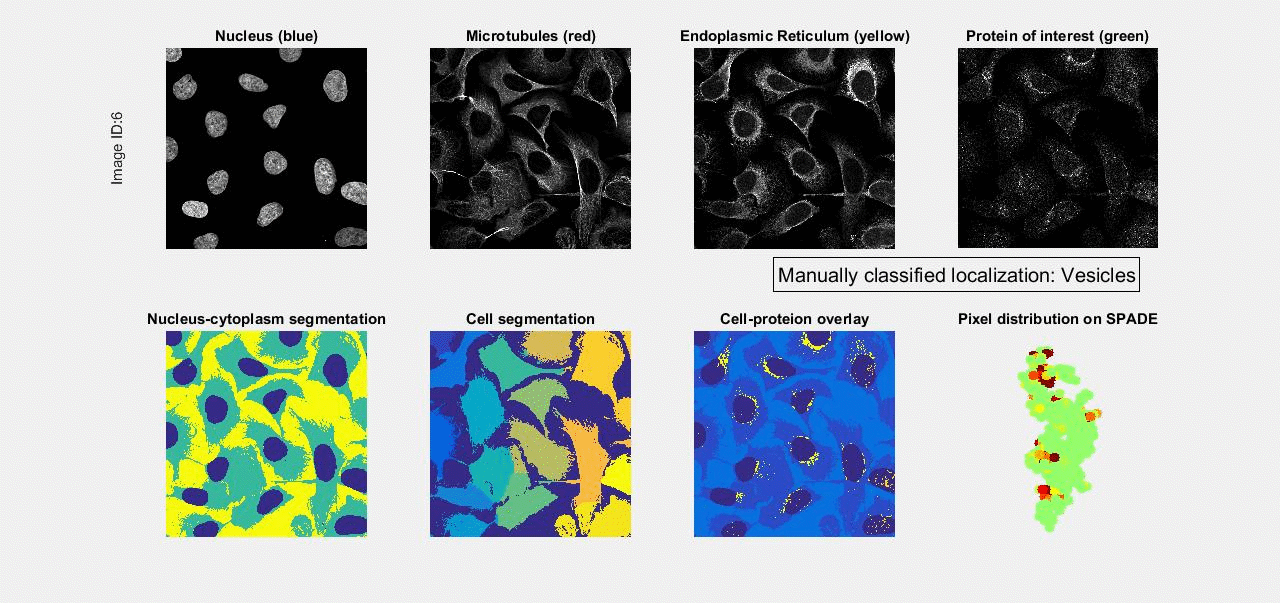
Below are examples of protein localizations that belong to 13 major classes (subcellular components), obtained from
the CYTO 2017 Image Analysis Challenge. For each of the 13 classes, 20 examples are provided. All these examples are
manually labeled with only one of the 13 classes. In the challenge, more than half of the examples are more complex,
and are labeled with multiple classes. From these relatively not-too-complex examples, we can see the varibility within
each class, as well as similarity/confusion among different classes. In addition to the mutli-class examples, another
complicating factor is the heterogeneity among cells in the same field-of-view, not all cells in the same image exhibit
the same localization.
In each example, the four gray-scale images in the top row are the raw data. Biology experts visually inspected these
four images to determine the localization of the protein of interest. The manually/visually determined localization
can be one or several of the 13 classes. The bottom row shows results of our analysis when participating in the challenge,
visualizing results of our image segmentation, thresholding, and SPADE-based feature extraction.